
Parkinson’s disease is a disorder that affects movement, and detecting it early remains a clinical challenge.
Thanks to advanced techniques such as PET/CT in Parkinson’s disease, today it is possible to identify changes in brain dopaminergic function even before the most obvious symptoms appear.
In Parkinson’s disease there is a progressive degeneration of dopaminergic neurons.
This involves a decrease in dopamine, alterations in movement control and the progressive appearance of symptoms such as tremor, rigidity or slowness.
These changes may begin years before the patient notices clear symptoms. PET/CT in Parkinson’s disease is therefore particularly useful in the early stages.
PET/CT in Parkinson’s disease is an advanced imaging test that combines functional and anatomical information.
On the one hand, PET makes it possible to assess the metabolic activity of the brain. On the other hand, CT provides detailed structural images.
This combination not only allows us to study the anatomy of the brain, but also how it functions. In the case of Parkinson’s disease, PET/CT enables precise analysis of the dopaminergic system.
PET/CT in Parkinson’s disease uses a specific tracer called F-DOPA that binds to dopaminergic systems in the brain.
It enters dopaminergic neurons, is converted to dopamine and enables measurement of dopaminergic synthesis and storage capacity.
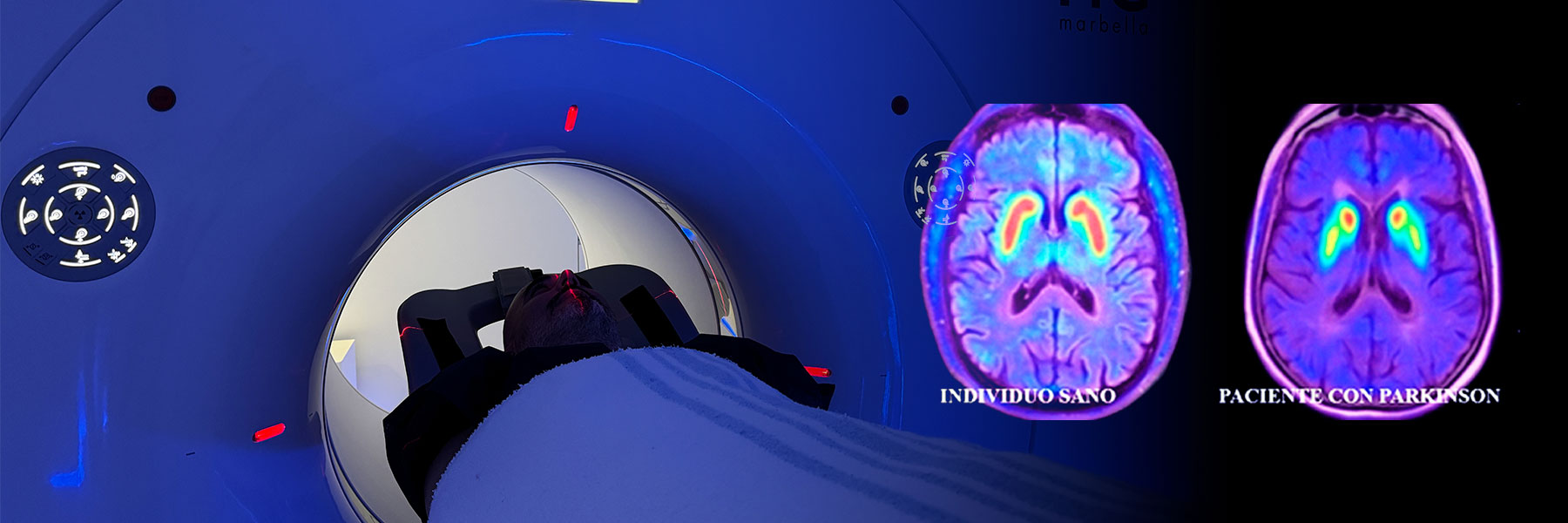
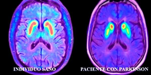
In people with Parkinson’s disease, a decreased uptake of this tracer is observed, especially in regions such as the striatum.
This shows the loss of dopaminergic neurons and makes it possible to detect abnormalities even in the early stages of the disease.
PET/CT in Parkinson’s disease can be useful in different clinical situations, especially when the diagnosis is inconclusive.
It is recommended for patients with uncertain initial symptoms, in cases of differential diagnosis with other similar disorders (atypical parkinsonian syndromes or Parkinson-like conditions), patients with atypical evolution or to assess the response to treatment.
Although it is not necessary in all patients, PET/CT in Parkinson’s disease provides key information in certain specific situations.
PET/CT in Parkinson’s disease is a simple, safe and well-tolerated test.
The tracer is first administered intravenously. Then, a waiting period allows it to distribute throughout the body.
Finally, the patient lies down on the imaging equipment for a few minutes to obtain the corresponding images.
It is painless and does not require hospital admission.
PET/CT in Parkinson’s disease allows for the early detection of changes in dopamine metabolism.
This significantly improves the management of the disease. It facilitates a more accurate diagnosis, allows earlier initiation of treatment and helps to plan a more appropriate follow-up care.
Furthermore, it reduces the patient’s uncertainty.
PET/CT in Parkinson’s disease is a key tool in the modern approach to the disorder.
It allows direct visualisation of changes in brain structures involved in movement control.
Its use in selected patients contributes to an earlier, more accurate and personalised diagnosis.
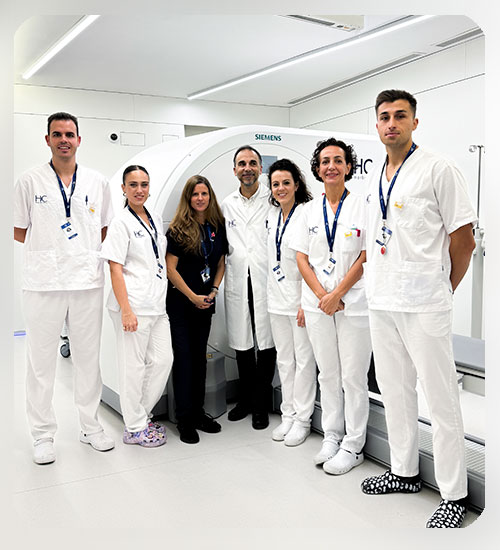
If you need a specialised assessment or have symptoms compatible with Parkinson’s disease, at HC Marbella we have state-of-the-art technology in nuclear medicine and a team of experts in neurological diagnosis.

Dr. Luis Villar
Doctor specialising in Nuclear Medicine
April 10, 2026
lesen Sie Nachrichten
Tel.: +34 952 908 628
+34 609 148 799
952908898 Onkologie
951829978 Bildgebende Diagnostik
951829947 Gynäkologie
952908897 Fertilitäts-Zentrum
951829947 Physiotherapie