Dr. Hernández Rubiño, Antonio
Otorrinolaringología
Jefe del servicio
Especialista en otología

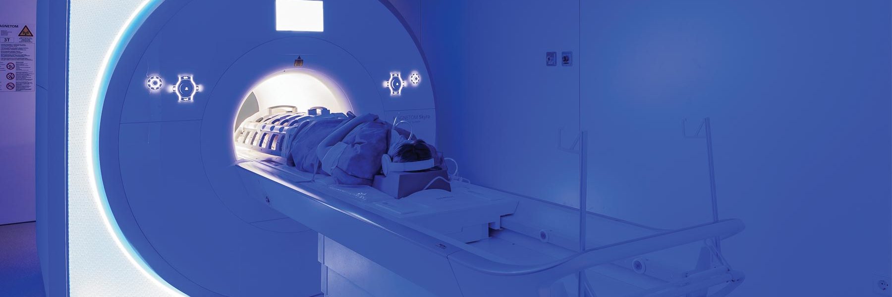
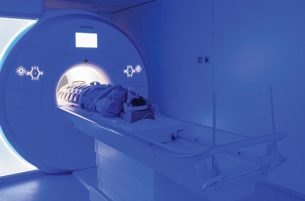

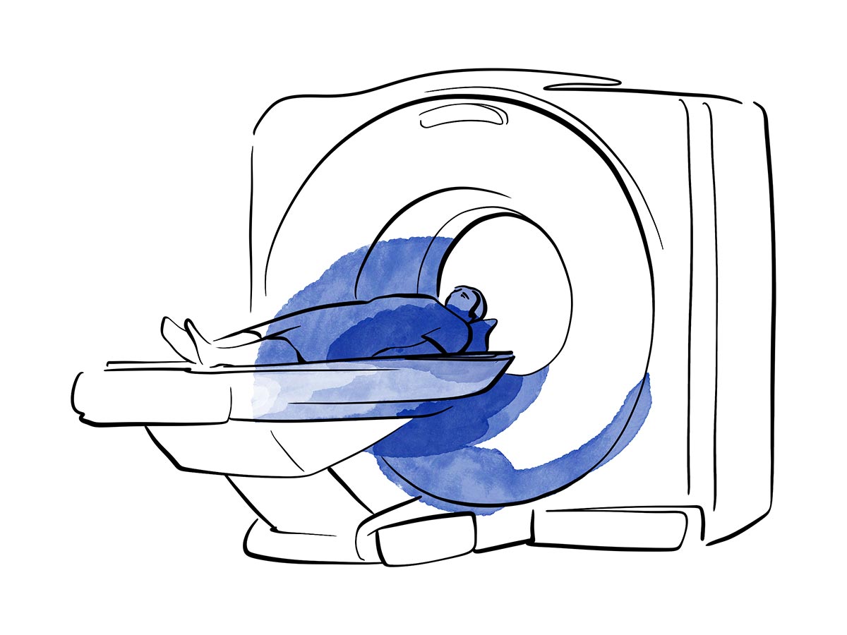
We offer the most advanced diagnostic tests, such as 3 Tesla magnetic resonance imaging and PET-CT, which allow precise detection and correct therapeutic planning.
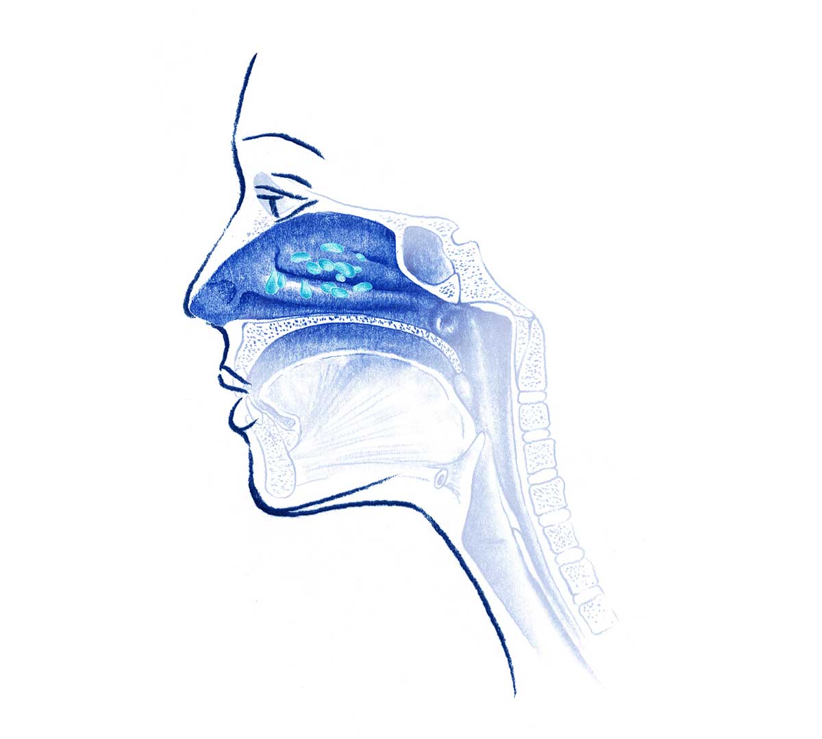
Nasofibrolaryngoscopy is a diagnostic technique in which the otolaryngologist inserts a thin, flexible endoscope equipped with a light and camera through the nose to examine the nasal cavities, pharynx and larynx (including the vocal cords) directly and in real time.
This procedure allows visualisation of structural or functional alterations of the upper airway (such as inflammation, tumours or changes in the mobility of the vocal cords) without the need for surgery. It facilitates both visualisation of the lesion and the performance of a guided biopsy.

FNA (Fine Needle Aspiration) is a procedure that involves inserting a thin needle into the suspicious lesion to extract cells or small pieces of tissue. This material is then analysed by the pathologist to determine if malignancy is present or to indicate the type of tumour.
It is a quick, minimally invasive procedure and is usually performed under ultrasound guidance to increase accuracy and avoid complications.
Magnetic Resonance Imaging in the diagnosis of head and neck cancer allows for the clear visualisation of both the primary tumour and the extent of its local and lymphatic spread, thanks to its excellent contrast in soft tissues.
It is often used to diagnose tumours of the nasopharynx, oropharynx, base of tongue, salivary glands.
Contrast-enhanced MRI offers the following possibilities:
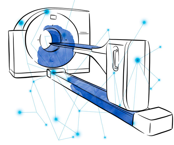
This diagnostic test is especially used in the diagnosis of laryngeal tumours or when there are contraindications to the use of magnetic resonance imaging.
It allows for the determination of tumour extension and the presence of adenopathies. Furthermore, it helps in the planning of treatment, as well as in the follow-up after treatment, by detecting recurrences or assessing complications.
It has a good capacity to show bone structures (such as the base of the skull) and for verifying the invasion of bone or cartilage.
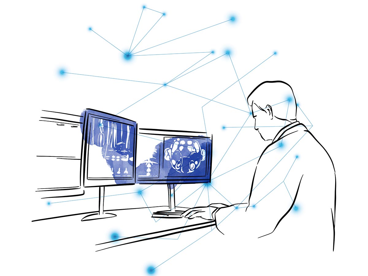
PET-CT (Positron Emission Tomography fused to Computed Tomography) is the most advanced and recommended imaging test for the management of head and neck cancer in complex cases. It combines metabolic information (PET) with high-resolution anatomical information (CT) in a single session and is indicated when there is suspicion of disease spread.
It has consolidated as a fundamental tool for diagnosis, cancer staging, treatment planning and evolutionary control or patient follow-up.
PET-CT is the test of choice for:
Dr. Hernández Rubiño, Antonio
Otorrinolaringología
Jefe del servicio
Especialista en otología

Dr. Tenor Serrano, Rafael
Otorrinolaringología
Especialista en cabeza y cuello
Apnea del sueño

Dr. Salvatierra Vicario, Belén
ENT
Specialist in endoscopic sinus surgery

Dr. Chiti-Batelli, Sandro
Otorrinolaringología
Especialista en rinoplastia estética y funcional y
cirugía endoscópica de senos paranasales

Dr. Rosas Marqués, Paloma
Audiometrías y pruebas funcionales
Tel.: +34 952 908 628
+34 609 148 799
952908898 Onkologie
951829978 Bildgebende Diagnostik
951829947 Gynäkologie
952908897 Fertilitäts-Zentrum
951829947 Physiotherapie