Dr. Herrera Ruiz, Marco Antonio
Odontólogo General y Prostodoncista

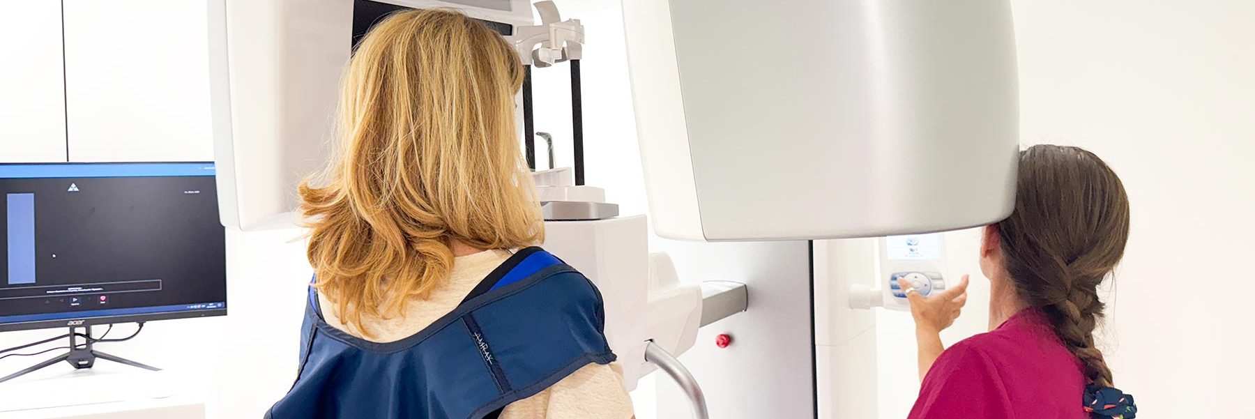
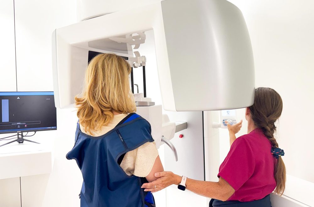
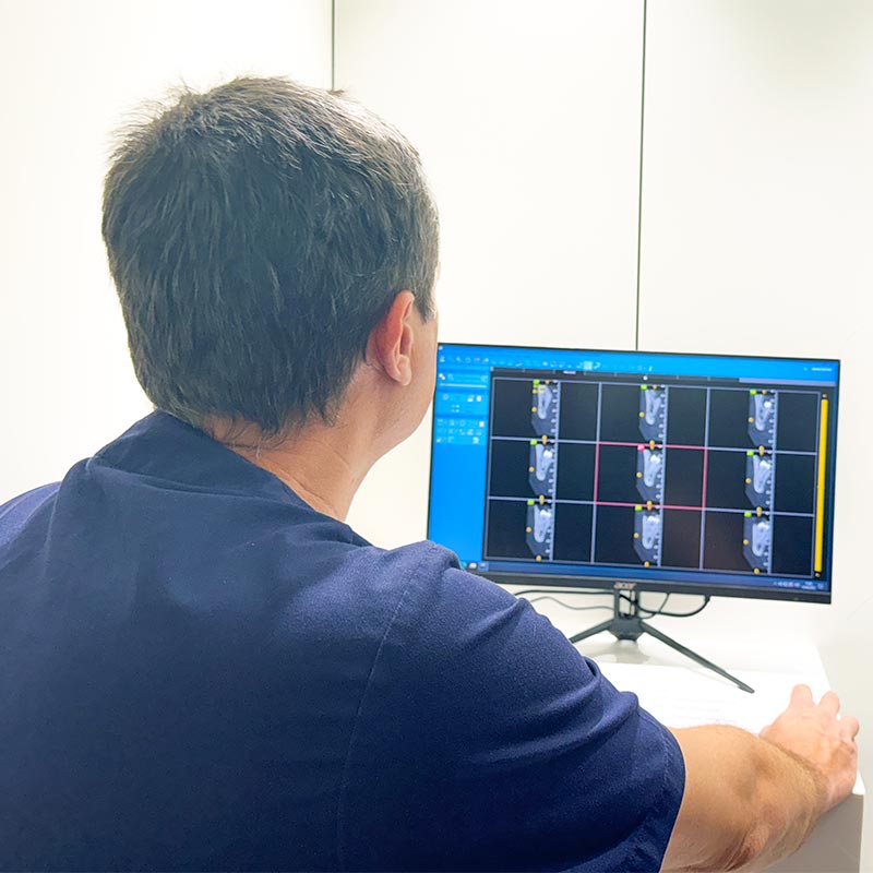
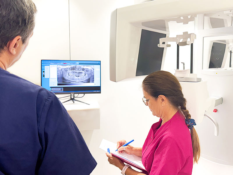
A revolution in dental diagnosis and treatment.
High-quality 2D and 3D images
Volumetric, panoramic, teleradiographic and dynamic radiographic examinations.
Lower radiation dose
Thanks to the ECO Scan mode, the radiation dose is considerably reduced.
Versatile and effective device
multiple selectable dimensions and different scan modes, this enables clear definition of the field of view to be studied.
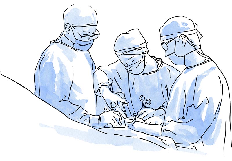
Complete view of the entire maxillofacial area, with maximum precision with regard to anatomical details. Ideal for oral and maxillofacial surgery procedures, it allows surgeons to clearly identify the presence of impacted teeth or fractures, assess bone density and height, as well as the morphology and orientation of dental roots.
Image quality is not compromised by the presence of metallic materials; the low dose of radiation emitted significantly reduces scattering, facilitating clear visualisation of anatomical structures.
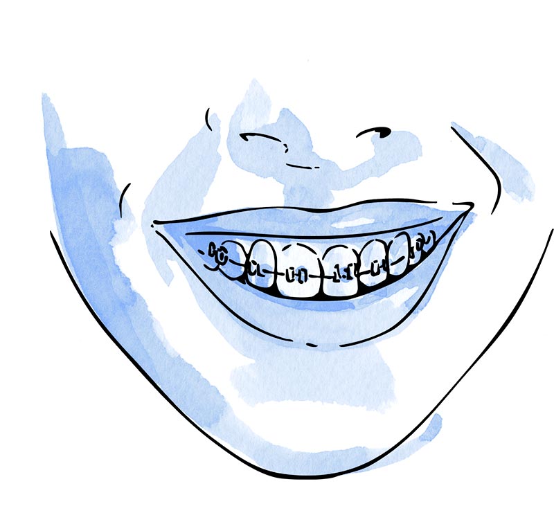
Cone Beam technology is particularly useful in orthodontic treatment for aesthetic or functional purposes. The three-dimensional images captured represent the area being scanned very clearly and in great detail, generating panoramic, teleradiographic and 3D images.

3D volumetric reconstructions reveal with high precision the presence of pathologies or anatomical alterations.
The use of Cone Beam technology allows detailed scans for precise implantological planning, evaluating the insertion site, bone width and the anatomical characteristics of the implant.
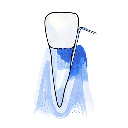
This technology allows the acquisition of sharp and detailed images for endodontic and periodontal studies, facilitating the detection of fractures, analysis of the mandibular canal and evaluation of adjacent tissues.
Such diagnostic accuracy allows for clear identification of any abnormalities, precise identification of the pathology and effective planning of the most appropriate treatment.
There are multiple study modes depending on the different clinical applications. A wide range of fields of view (FOV) can be selected, determining the extent of the anatomical region analysed.
Wide fields of view facilitate capturing complete images of facial bone structure in a single scan, ideal for orthodontic, orthognathic and maxillofacial surgery procedures.
Medium fields of view are suitable for temporomandibular joint (TMJ) assessment, full denture analysis and implant treatment planning.
Small fields of view are particularly suitable for endodontic, periodontic and implantology studies in localised areas, adapted to the needs of the patient.
Dr. Herrera Ruiz, Marco Antonio
Odontólogo General y Prostodoncista

Pérez Bocanegra, Marta
Ortodoncista

Gorospe, Adriana
Implantóloga

Doblas López, Carmen Marina
Higienista Dental

Bustos Solero, Claudia
Higienista Dental
Tel.: +34 952 908 628
+34 609 148 799
952908898 Oncology
951829978 Diagnosis by imaging
951829947 Gynecology
952908897 Fertility
951829947 Physiotherapy