Dr. Ciano Petersen, Nicolás Lundahl
Neurologist at HC Marbella

The more accurate the diagnosis, the better the choice of treatment.
The more we know about a disease, the better we can treat it and the more effectively we can help the patient.
At HC Marbella International Hospital we provide:

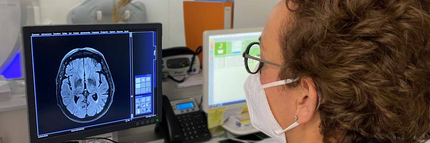
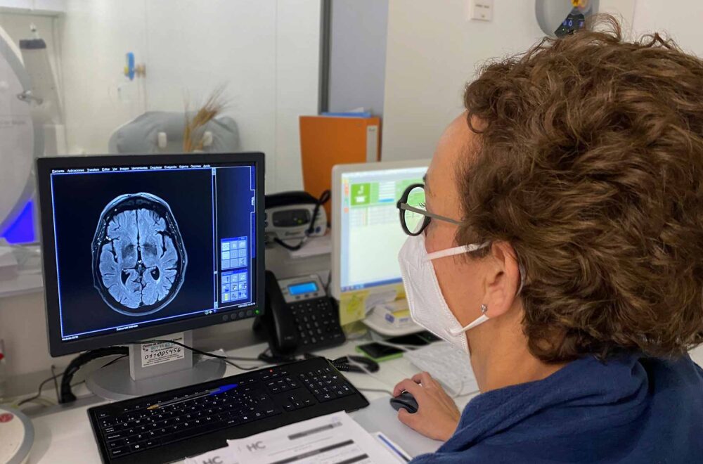
In neurology there are two reasons to order neuroimaging tests:
Types of neuroimaging tests:
Advanced neurophysiology tests allow the study and real-time analysis of the functionality of the central and peripheral nervous system to provide essential information relevant to the diagnosis and evolution of several neurological diseases.
The Neurophysiology studies available at the HC International Hospital in Marbella are:
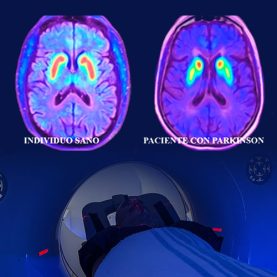
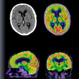
PET is a fundamental diagnostic technique in neurological disease. Anatomical/metabolic information can be obtained during a single procedure, in real time, increasing diagnostic accuracy. The technique combines:
In neurology it is used for:
This is used to detect the presence of amyloid protein that accumulates in the brain of patients with Alzheimer’s disease and other dementias. It enables early detection of the disease, since the onset of amyloid accumulation begins up to 20 years before the onset of symptoms.
Using a specific tracer, it identifies accumulation of the Tau protein. In contrast to amyloid, which accumulates widely throughout the brain, the Tau protein is concentrated precisely where brain atrophy is most severe.
It shows increases or decreases in brain metabolism in numerous neurological diseases and psychiatric disorders. It facilitates early and differential diagnosis of dementias.
This provides high diagnostic accuracy in conditions affecting the central nervous system (brain, stem and neuro-axis).
Exceptional anatomical detail, especially in Neurology and Neurosurgery.
Indicated in:
Estudio tridimensional prequirúrgico de alteraciones del cráneo
These allow us to extract information from samples (blood, cerebrospinal fluid…) which make it possible to detect a disease at an early stage or to know how far along in the disease the patient is.
Specific biomarkers exist for:
This consists of recording the bioelectrical activity of the brain whilst awake or asleep and during distinct functions of brain activity (photostimulation, hyperventilation).
It is used to assess the level of consciousness, neurodegenerative disease and the presence of epileptic phenomena.
This records electrical activity of the peripheral nervous system. It involves sub-tests: an electroneurogram by which nerve conduction velocity is measured, and electromyography which analyses the neurophysiological activity of the muscle.
It is used to rule out or confirm diseases such as Motor Neurone Disease, Polyneuropathy, Myasthenia gravis, Myopathies, Radiculopathies, etc.
This examination evaluates the function of the visual, auditory and somatosensory systems and their pathways, by means of response to a known and standardized stimulus.
Its results can rule out or confirm diseases such as Multiple Sclerosis, optical and auditory neuropathies, spinal cord injuries, etc.
This is the study of sleep and involves the comprehensive analysis of multiple simultaneous functions: Electroencephalography, Electromyography, Respiratory Function by measuring oxygen saturation and electrocardiography.
It is employed in the study of diseases that can affect sleep: sleep apnoea/hypopnoea syndrome, narcolepsy, insomnia, restless leg syndrome, etc.
Lumbar puncture consists of the removal of a small amount of cerebrospinal fluid. To enable this, the patient is placed in the left lateral position whilst lying down, with the knees bent up as close to the body as possible.
The fluid is tested for evidence of infection, cerebral haemorrhage, multiple sclerosis, metabolic disease or other neurological conditions.
Dr. Ciano Petersen, Nicolás Lundahl
Neurologist at HC Marbella


Tel.: +34 952 908 628
+34 609 148 799
952908898 Oncology
951829978 Diagnosis by imaging
951829947 Gynecology
952908897 Fertility
951829947 Physiotherapy