

Update yourself on the latest developments!
An ultrasound provides images of internal organs which have been constructed using sound waves. These images are produced by a probe (transducer), which when placed directly onto the skin sends ultrasound waves into the body. It then receives the waves which rebound off the organs, analysing them and constructing an image of the organs afterwards.

There are different types of ultrasound and different uses, advances over time have enabled higher quality images. In this article we will analyse their use in obstetrics, gynaecology and fertility specifically and we will discuss the latest technological advance which provides more realistic and vivid images of the baby in the uterus: 5D ultrasound.
5D ultrasound, a huge leap forward
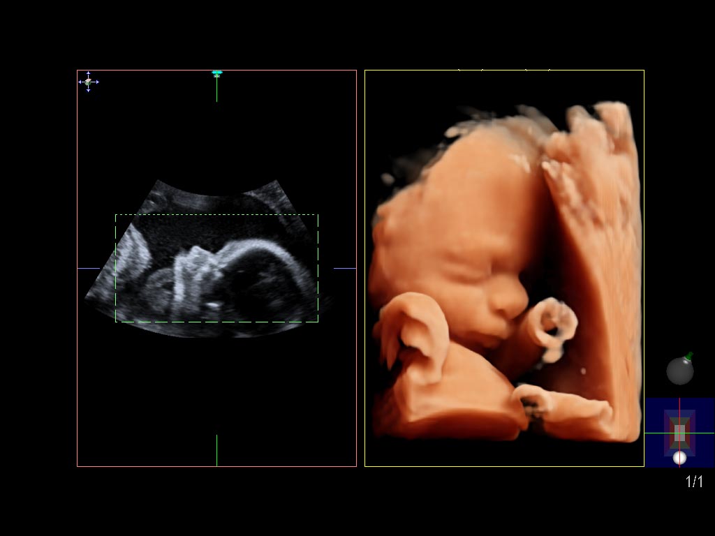
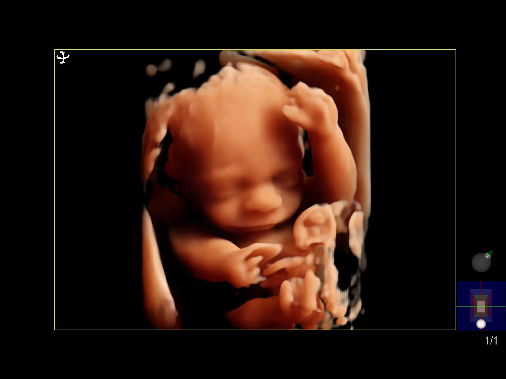
“Today we have new ultrasound equipment providing 5D ultrasound. This advance enables us not only to view the baby in the uterus before birth, with its movements in real time (the same as with 4D ultrasound) but also to better define the baby’s profile whilst establishing skin tones and generating more realistic, vivid images. This technology also allows us to more easily eliminate interference between the probe and the face of the baby, thus providing us with a more accurate picture, giving the image clarity and volume. Innovation in this field includes the incorporation of VSI (Volume Shading Imaging) technology. It produces improved visualisation of superficial anatomical structures, achieving skin tones and shading which generate so-called “natural” images, in the same way as 3D imaging. They are similar to those obtained via fetoscopy, a technique which allows visualisation of the foetus, placenta and umbilical cord,” explains Dr Pakzad, Consultant Gynaecologist and Obstetrician at HC Marbella. He continues,
“The latest generation ultrasound using 3D technology allows evaluation of certain structures within the baby, such as the brain or spine. It also allows evaluation of the uterus or ovaries in patients who are not pregnant, providing three dimensional images, complementing conventional 2D ultrasound in the diagnosis of certain specific conditions. Measurements can be taken afterwards as details of the volume can be stored and reviewed as often as required.”
This type of imaging has changed the way we can see the baby inside the uterus. However Dr Pakzad explains that it is not the only use for this type of technology. “This new technology is of high resolution and able to assess blood flow using colour Doppler, fundamental in the investigation of different haematological and immunological conditions.”
Ultrasound during pregnancy allows monitoring and detection of different abnormalities from the first trimester onwards, however it can also play an essential role in the second and third trimesters. Regarding 5D ultrasound, Dr Pakzad explains, “The ideal moment to have a 5D ultrasound is the same as for a 4D scan, between 26 and 29 weeks, the baby is then the ideal size to be seen and the amount of amniotic fluid at this time is sufficient to obtain good images. However the baby always needs to be in an appropriate position to be viewed, not hidden by arms or legs, or the placenta, with a normal quantity of amniotic fluid, etc. During the first trimester the images of the embryo or intrauterine environment are truly sharp, it is possible to see the whole uterine cavity, showing the relationship between the two.” This is a revolution in the field of maternal-foetal medicine. It has not only had a medical impact, but it has also given society the opportunity to establish a much deeper emotional connection with newborns than ever imagined.
Whilst ultrasound is the “star” during pregnancy, it also plays a very important role from the gynaecological point of view, including in cases of infertility.
“3D ultrasound is a transvaginal ultrasound which allows us to see the uterus as a whole, obtaining different slices to those obtained with common, or 2D, ultrasound. This facilitates the diagnosis of certain conditions which can affect fertility such as: uterine malformation, adhesions between the walls of the uterus, fibroids which grow inside the uterus, adenomyosis etc.,” explains Dr Pakzad, giving another detailed example: “The hormone releasing IUD for example is often more difficult to see with ultrasound due the material from which it is made. Generally we see the shadow which it makes, different to the conventional copper IUD which is bright. When we use transvaginal 3D ultrasound we can obtain very clear images of the location of the shadow which the hormone releasing IUD generates, allowing us to see its position very clearly.
“In the case of adenomyosis, a benign condition of the uterus, glands making up the endometrium invade the muscle walls of the uterus, increasing its size and, amongst other things, altering its receptivity. Ultrasound is fundamental in this condition as it is often confused with fibroids, common benign tumours. Fibroids do not usually cause problems, except when they are very large or alter the uterine cavity, compromising implantation of an embryo. A fibroid may require surgery if it interferes with attempts to achieve pregnancy, alternatively adenomyosis, although it does not cause serious problems, does not provide this possibility. In some cases it can reduce implantation rates, hence the importance of its diagnosis.”
Dr Pakzad also goes on to talk about the use of sonohysterography. “This is an ultrasound which is performed after inserting a contrast solution into the uterus, through a very fine catheter measuring around 1 mm. This study is used to assess the uterine cavity and enables the diagnosis of polyps, fibroids and uterine synechiae, conditions which can affect the implantation of an embryo. We can combine it with 3D technology to obtain improved images, even evaluating them once the patient’s test has finished.
Both during pregnancy and when trying to achieve pregnancy, the latest technological advances in ultrasound have a great contribution to make. In trained hands ultrasound is an incredibly useful diagnostic too.
We have the latest generation ultrasound equipment at HC Marbella, including 5D for use according to patients’ needs and preferences. Come to meet us, we will happily provide you with any information you require, with no obligation.
February 6, 2018
Read other news
Tel.: +34 952 908 628
+34 609 148 799
952908898 Oncology
951829978 Diagnosis by imaging
951829947 Gynecology
952908897 Fertility
951829947 Physiotherapy


