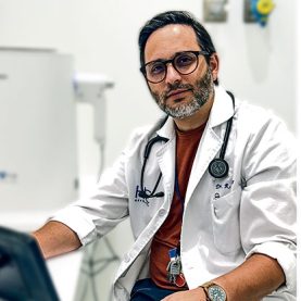
Dr. Aguilar PerezGrovas, Ricardo
Neumology Specialist

Lung biopsy is a diagnostic procedure that allows us to look for suspicious tissues in the airways and lungs and to obtain samples of this tissue for further analysis in the laboratory.
This procedure can determine whether there is any lung cancer or lung disease.
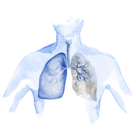
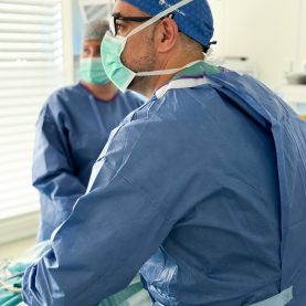
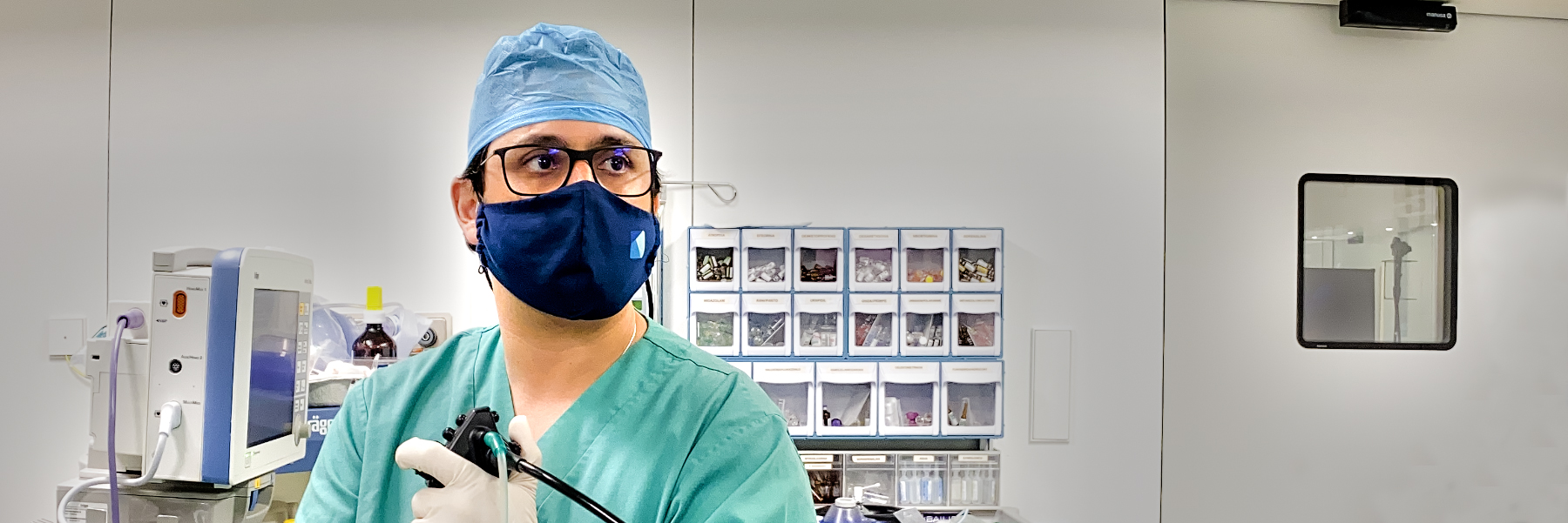
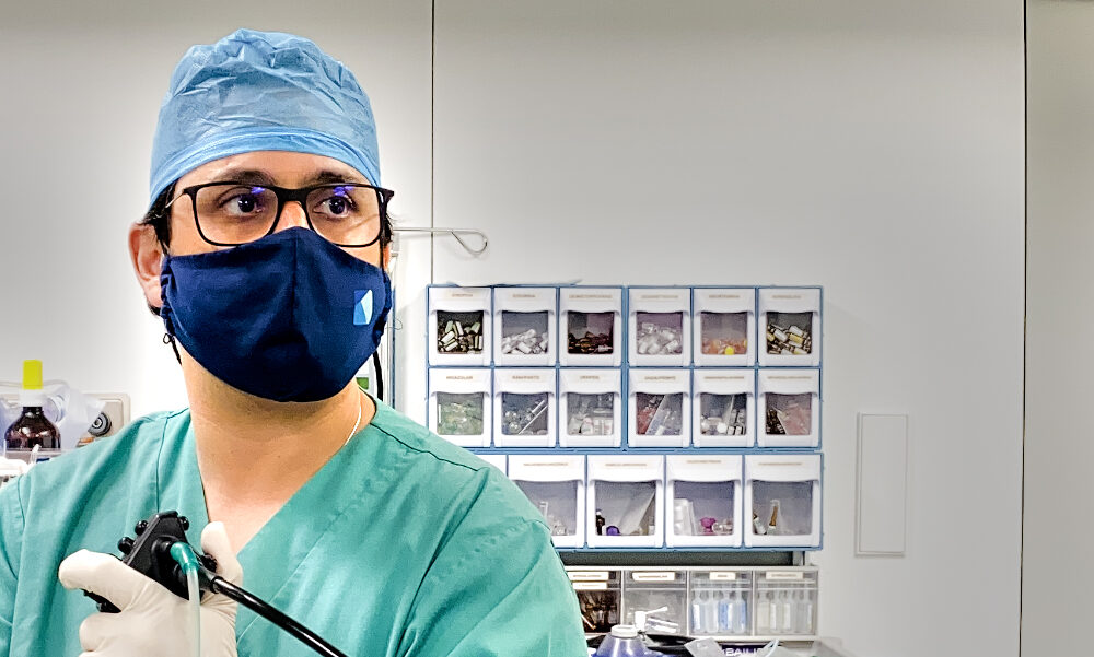
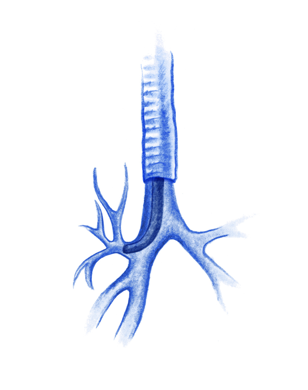
Bronchoscopy is used to observe airway abnormalities using a bronchoscope – an endoscopic tube with a camera at the tip – it is inserted through the nose or mouth to the airways in the lungs.
When is it indicated?
It is used as a secondary test for those patients who require a biopsy to diagnose the cause in the case of suspected bronchopulmonary tumours, lymphadenopathy, metastatic tumours and/or infectious conditions, such as severe pneumonia, bronchial secretions or suspected tuberculosis.
How is this performed?
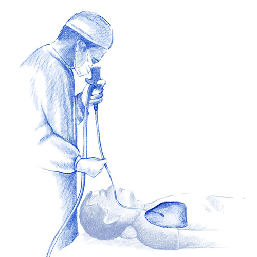
The test is performed using topical anaesthetic spray and sedation to make it as comfortable as possible for the patient. After the patient has been sedated, the bronchoscope is inserted through the nostril and the upper and lower airways are explored
The duration of the test varies, depending on the indication and the findings it can be from 15 to 45 minutes. The procedure is performed in an outpatient setting, the patient may go home within a few hours after recovering.
Mediastinoscopy is a procedure used to view the mediastinum. This is an area in the centre of the chest, between the lungs and where lymph nodes are usually present along with the veins and arteries. A mediastinoscope, a thin tube with a light and a lens, is used to do this. Sometimes forceps are introduced through the tube to remove a tissue sample which is then analysed for signs of disease.
When is it indicated?
This test is indicated in people when suspicious tissue or abnormal lymph nodes have been found on CT scan of the chest, generally in inflammatory or infectious diseases, bronchopulmonary or haematological malignancies.
How is it done?
It is performed under general anaesthetic. After a small incision is made in the upper sternum, the mediastinoscope is inserted into the middle of the chest. Thanks to the camera in the tube, the specialist can observe the mediastinum. If suspicious tissue is observed, a sample of the tissue or lymph nodes around the lung can be removed
A chest X-ray may be performed after the test.
Lung or pleural biopsy provides lung or pleural tissue for sampling or the diagnosis of diseases such as cancer or severe infection. It is performed when an abnormality has been detected by an imaging test, usually a CT scan or chest X-ray.
Tissue samples are then analysed in the laboratory.
There are different procedures for performing the biopsy, each case is different, it may be transbronchial, puncture, via thoracoscopy or by open biopsy.
In most cases, lung biopsy can be performed on an outpatient basis or as part of a hospital stay. It is carried out using a mild local anaesthetic to numb the area.
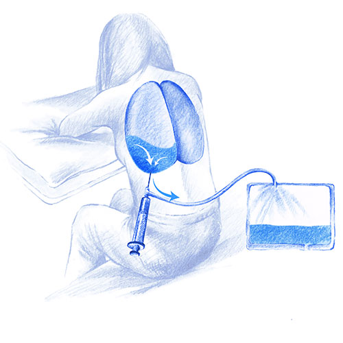
This test enables the extraction of pleural fluid for diagnostic purposes or as part of treatment (e.g. pleural effusion in patients with renal failure who do not respond to medication or dialysis, or patients with malignant lung disease).
Normally, the pleural cavity contains a very small amount of fluid. This procedure is performed to drain fluid from the space between the outer lining of the lung (pleura) and the chest wall.
How is it done?
Extraction is performed under local anaesthetic. A needle is injected through the skin which then collects the fluid to be sent for laboratory analysis (pleural fluid analysis).
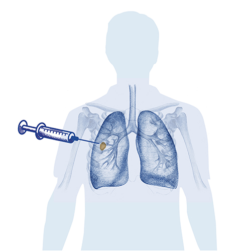
Transthoracic puncture is a diagnostic procedure in which a fragment of lung tissue is removed for further analysis.
It is an outpatient test and usually lasts 30 to 60 minutes.
To take the sample, the interventional radiologist inserts a needle through the chest wall into the suspected tissue and a small sample of tissue is removed.
The procedure is usually guided by a CT scan or chest X-ray to locate the lesion to be biopsied.
Bronchoalveolar lavage is a procedure in which samples of cells are taken from inside the airways that lead to the lungs for further analysis.
When is it indicated?
This test is usually carried out to diagnose different infections such as tuberculosis, bacterial pneumonia, or fungal infections. It sometimes helps to detect cell changes that can lead to lung cancer.
How is it done?
It is carried out by inserting a bronchoscope through the nose or mouth into the lungs. A saline solution is introduced through this in order to wash out the airways and collect cells that will then be analysed under the microscope.
Dr. Aguilar PerezGrovas, Ricardo
Neumology Specialist
Tel.: +34 952 908 628
+34 609 148 799
952908898 Oncology
951829978 Diagnosis by imaging
951829947 Gynecology
952908897 Fertility
951829947 Physiotherapy