
New advances in the treatment of triple-negative breast cancer

Our priority is to provide an accurate diagnosis and the most appropriate treatment in each case.
To this end, our gynaecologists work with the latest ultrasound systems and in close collaboration with the diagnostic imaging unit and laboratory, both located in the same area of the hospital and equipped with the most advanced technology.
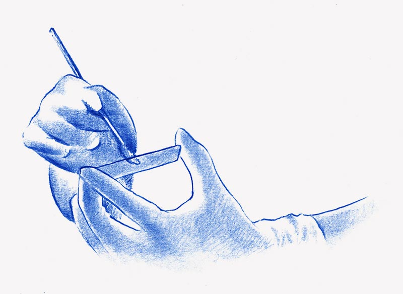
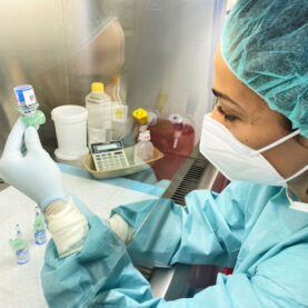
Cytology is a routine test included in your annual gynaecological check-up, it is also known as a Pap smear. This is a simple and quick technique, where cells from the vagina and cervix are gently scraped to obtain the specimen.
The goal of this test is to detect lesions that are precursors of cervical cancer or cervical cancer itself. It also enables the diagnosis of infections and is sometimes used to obtain a hormonal diagnosis.
An abnormal cytology result does not necessarily involve a cancerous lesion, other conditions such as infections, herpes, trichomoniasis, etc. may be the cause. In such cases, treatment or monitoring will be required.
Depending on the scale used, the interpretation of cytology results may be:
Classic Method:
A different scale:
“TBS” classification:
Depending on each case, the gynaecologist will assess the potential causes of the abnormality and recommend the most appropriate treatment.
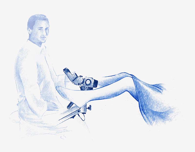
Colposcopy involves an amplified inspection, by means of a magnifying microscope, of the cervix, vagina (vaginoscopy), and vulva (vulvoscopy), it is carried out directly and with the use of filters and reagents.
It enables:
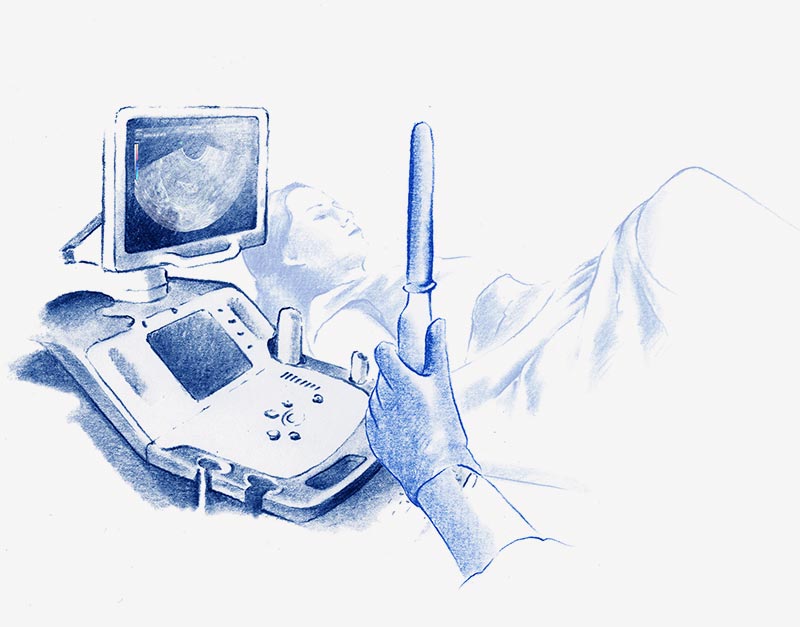
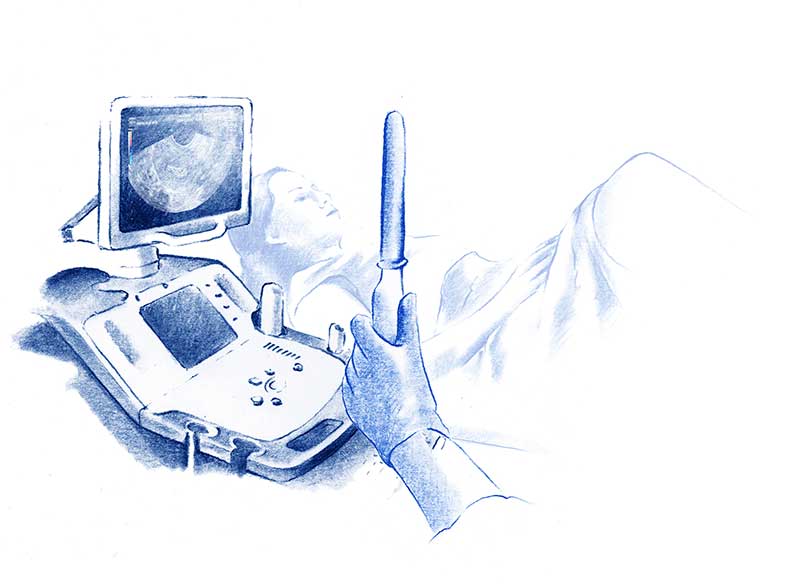
Like cytology, transvaginal ultrasound is a common test included in your annual gynaecological check-up.
Using an ultrasound probe, the female reproductive organs: uterus, cervix, vagina, ovaries, and Fallopian tubes can be fully examined. It allows the diagnosis and monitoring of pathological processes such as myomas, uterine malformations, infections, cysts, tumours, adhesions between the walls of the uterus etc. It can also determine the cause of bleeding or pelvic pain.
Ultrasound is also used when taking biopsies, it enables visualisation of the exact area from which a sample should be taken.
A number of state-of-the-art ultrasound systems are available at HC, offering exceptional image quality, enhanced colour sensitivity and advanced imaging modes.
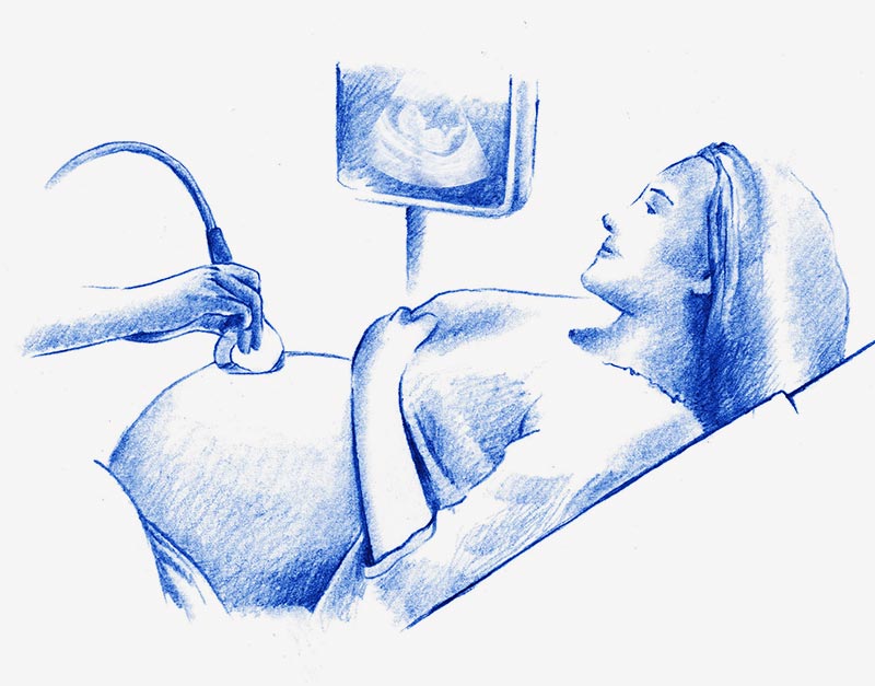
We currently use 5D-ultrasound systems, which allow us not only to see the image of the baby in the womb before birth, plus its movements in real time (as in 4D ultrasound), but it also intensifies outlines and establishes tones of shading on the skin, generating more vivid, realistic images. In addition, this technology enables us to more easily eliminate the interference between the probe and the face of the baby, giving a more accurate view, providing clarity and volume, being able to visualize the foetus, placenta and umbilical cord in a very detailed way.
This breakthrough in image quality has not only delivered increased diagnostic capacity, but it has also helped parents to have a reliable image of their child before birth, reinforcing the emotional bond.
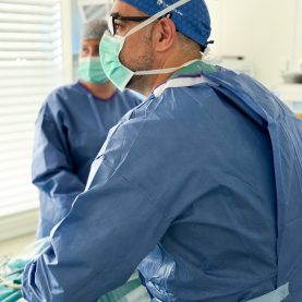
Diagnostic hysteroscopy is a relatively simple test, well tolerated and with few complications. It provides visualisation of the cervical canal and the uterine cavity, being a key study in the morphological and functional examination of the endometrium.
Hysteroscopy is usually indicated in the following cases:
The procedure is usually carried out as an outpatient and involves inserting the hysteroscope (lens) into the uterus, through the vagina, using a means of distension to separate the walls. A physiological or CO2 solution is also used to expand the uterine cavity. The lens allows direct images to be displayed on a monitor and recorded for further study, if necessary.
Hysteroscopy may use for surgery to perform biopsies, intervene in previously diagnosed pathology, or resect.
A routine, painless test performed at a gynaecological check-up or when the patient’s symptoms require it. This test consists of obtaining a sample of vaginal discharge using a swab, for later viewing under a microscope. It also enable culture of secretions to determine whether and which infection is present.
Dr. Escobar, Ángela
Ginecología y Obstetricia, especialista en la Unidad de Mama

Dr. Pakzad, Ramin
Ginecología y Obstetricia

Dr. Gerchunoff, José Ignacio
Finecología y obstetricia, especialista en la Unidad de Mama

Dr. Bizjak da Fonseca, Tina
Ginecología y Obstetricia, especialista en suelo pélvico

Dr. Sánchez Miguel, Tamara
Histeroscopia y Estudios de Fertilidad
Histeroscopia, Embarazo de Alto Riesgo y Estudios de Fertilidad

Dr. Mata, Jordana
Specialist in Human Assisted Reproduction

Dr. Utrera Jiménez, Almudena
Specialist in Human Assisted Reproduction

Dr. Lavezzari, Celina
Ginecología y Obstetricia
Dr. Avendaño García, Juan Diego
Ginecología y Obstetricia, especialista en Ginecología Oncológica

Dr. Rodríguez Rodríguez, Jose María
Ginecología y Obstetricia,
especialista en cirugía ginecológica y endometriosis
Tel.: +34 952 908 628
+34 609 148 799
952908898 Oncology
951829978 Diagnosis by imaging
951829947 Gynecology
952908897 Fertility
951829947 Physiotherapy