Dr. Jiménez Rodríguez, Begoña
Specialist in Medical Oncology
Clinical Dedication in Breast and Gynecological Cancer

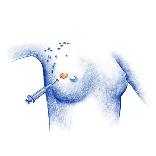
Breast biopsy is an interventional technique where samples of the lesion are removed and studied in the pathology laboratory to tell us exactly what type of lesion it is.
Biopsies may be performed at the time of surgery (such as sentinel node biopsy) or using image-guided puncture by the interventional radiologist on an outpatient basis.
Advantage:
Disadvantages:
This can be performed using ultrasound, stereotaxy, or magnetic resonance.
Procedure:
Advantages:
Disadvantages:
The sentinel node is the first lymph node to which cancer cells are most likely to spread from a primary tumour.
Procedure:
How is a sentinel node biopsy performed?:
Benefits:
Dr. Jiménez Rodríguez, Begoña
Specialist in Medical Oncology
Clinical Dedication in Breast and Gynecological Cancer

Dr. Sedano Ferreras, Paula
Radiotherapy Oncology Specialist

Dr. Subires Bootello, Marta.
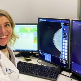
HC Marbella Radiology Specialist
Dr. Escobar, Ángela
Ginecología y Obstetricia, especialista en la Unidad de Mama

Dr. Rebollo García, Natividad
HC Marbella Radiology Specialist

Dr. Bellinvia, Anna Alessandra
HC Marbella Radiology Specialist

Dr. Gerchunoff, José Ignacio
Finecología y obstetricia, especialista en la Unidad de Mama

Dr. Martín Suárez, Luis
Specialist in the Plastic and Reconstructive Surgery

Dr. Arrazola, Tomás
Especialista en Farmacia Hospitalaria
Especializado en terapia contra el cáncer, certificado por la Sociedad Americana de Farmacéuticos de Hospital
Tel.: +34 952 908 628
+34 609 148 799
952908898 Oncology
951829978 Diagnosis by imaging
951829947 Gynecology
952908897 Fertility
951829947 Physiotherapy