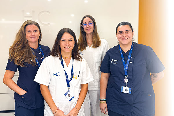

Dr Marta Subires joins the breast imaging team at HC Marbella

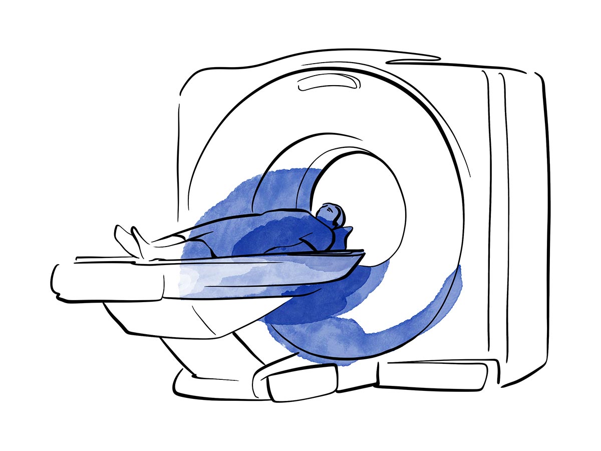
Breast MRI is an advanced imaging test that makes it possible to assess breast tissue in great detail and obtain information that mammogram or ultrasound cannot always provide on their own.
It can help to detect and characterise lesions, assess the extent of a cancer that has already been diagnosed, monitor response to treatment and evaluate the condition of breast implants.
At HC Marbella, we perform this scan using a 3-Tesla MRI scanner with artificial intelligence, which provides high-definition images in less time.
Breast MRI complements mammogram and ultrasound, but does not replace them. Also, it does not use ionising radiation.
Magnetic resonance imaging is an imaging test that uses a magnetic field and radio waves to create detailed images of the inside of both breasts.
In most scans aimed at detecting or assessing lesions, an intravenous gadolinium-based contrast agent is administered. This contrast agent allows us to assess tissue behaviour and evaluate any suspicious areas with greater precision.
Key features of the test:
Greater clarity. Less time.
Artificial intelligence optimises image acquisition, making the scan faster, more comfortable and more precise.
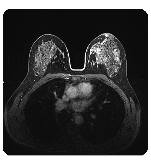
A breast MRI scan makes it possible to:
Women at high risk of breast cancer
It is recommended for women with:
Following a breast cancer diagnosis
When a biopsy confirms cancer, an MRI scan helps to determine:
Treatment monitoring
It allows for a comparison of images taken before and after treatment to assess how the tumour is responding, and helps the medical team plan the next steps with greater precision.
Findings requiring further investigation
Important note: in women at high risk, an MRI scan is usually carried out alongside a mammogram as part of a personalised monitoring programme; it does not replace it. The final decision is made by your specialist based on your medical history and previous test results.
Not sure which test you need? Find out more about breast diagnostic imaging.
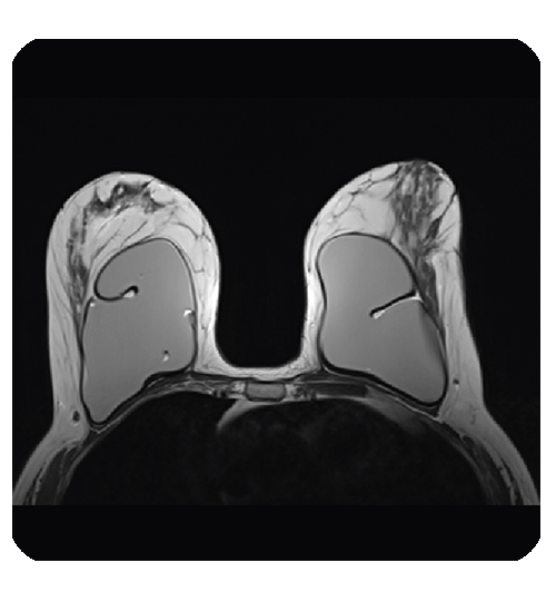
MRI makes it possible to check the integrity of silicone implants and detect any potential ruptures or leaks.
When the aim is solely to evaluate the implant, there is a specific protocol that usually does not require contrast. If it is also necessary to assess the breast tissue, a different protocol will be used.
The results are reported using the international BI-RADS classification system, which is also used for mammograms and ultrasounds.
Important note: the BI-RADS result is not a diagnosis in itself. A suspicious finding (BI-RADS 4 or 5) does not necessarily mean cancer; it indicates that further investigation is required, which may include an ultrasound, a biopsy, or ongoing monitoring. Your specialist will interpret the result alongside the rest of your test results.
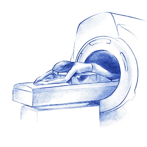
During the scan, you will lie face down on an examination table. Your breasts are positioned in specific slots that allow images to be taken without compressing them.
The test is painless, although it is important to remain still. In scans aimed at detecting or characterising lesions, an intravenous contrast agent is usually administered.
The duration depends on the required protocol. You will be able to communicate with the radiographer throughout the whole scan.

Before the test, it is essential to inform us if you:
For women who are still menstruating, it may be recommended that the scan be performed at a specific stage of the cycle.
It is also important to bring along previous mammograms, ultrasounds, MRI scans and reports so that the results can be compared.
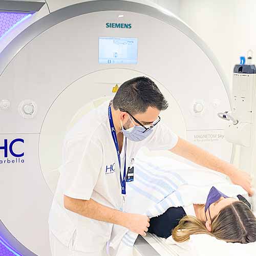
Do you need a breast MRI? Book an appointment with our Breast Unit.
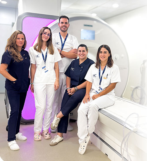
Technology makes an important difference, but it is our clinical team that turns it into a reliable diagnosis.
No. They are complementary tests that provide different information. The specialist will decide which tests are necessary in each case.
No. An MRI scan uses a magnetic field and radio waves.
No, it is painless and does not compress the breast. However, lying face down while remaining still may be slightly uncomfortable.
Tests designed to detect or assess lesions usually require contrast. However, when evaluating implants alone, a protocol without contrast may be used.
Yes. MRI can be used both to assess breast tissue and to evaluate the condition of silicone implants.
Not necessarily. MRI can also reveal benign abnormalities. The radiologist will advise whether monitoring or further investigation is required.
Yes. Comparing current images with previous tests helps to interpret the findings more accurately.
A 3T breast MRI provides detailed imaging to support diagnosis, assess the extent of a lesion, or carry out personalised monitoring.
Do you need a breast MRI scan?
Book an appointment with our Breast Unit or the Diagnostic Imaging Department at HC Marbella.

De Castro, Francisco Javier
Medical specialist in Radiology and Internal Medicine
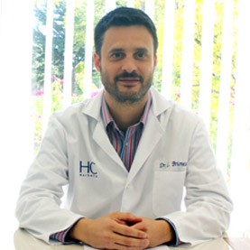
Briones, Luis
HC Marbella Radiology Specialist

Bellinvia, Anna Alessandra
HC Marbella Radiology Specialist

Rebollo García, Natividad
HC Marbella Radiology Specialist
Subires Bootello, Marta.
HC Marbella Radiology Specialist

Rivera Sánchez, Ernesto
HC Marbella Radiology Specialist
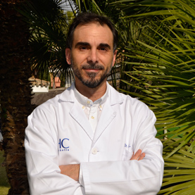
Villar Luque, Luis
Especialista en Medicina Nuclear

García Baltar, José Antonio
Specialist in Hospital Radiophysics and Radiation Protection
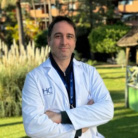
Cano Iglesias, Enrique
Specialist in Hospital Radiophysics and Radiation Protection

Urbaneja Salas, Alberto
Specialist in Interventional Radiology

Rueda Galiano, Mª Isabel
Senior Technician in Diagnostic Imaging
Tel.: +34 952 908 628
+34 609 148 799
952908898 Oncology
951829978 Diagnosis by imaging
951829947 Gynecology
952908897 Fertility
951829947 Physiotherapy