ecografia 4d
ecografia 4d

Ultrasound

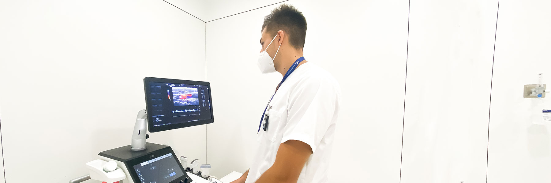
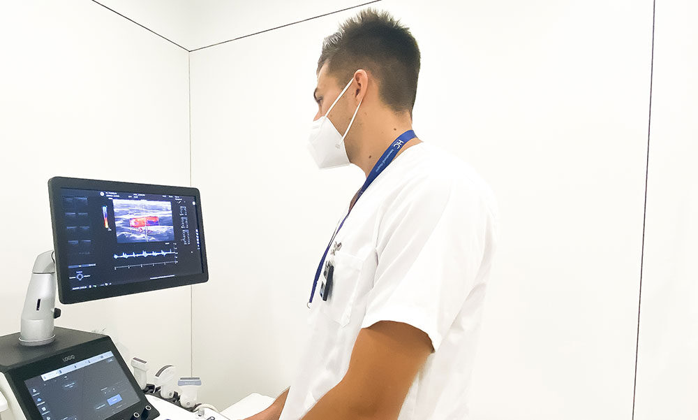
HC has several ultrasound machines designed for different uses.


We employ the latest innovations in technology to offer imaging of exceptional quality, with greater colour sensitivity and advanced imaging modes.


ecógrafo

How does it work?

Ultrasound is a diagnostic imaging technique which uses high-frequency sound waves to obtain images from within the body, different tissues reflect sound waves in varying ways, according (approximately) to their fluid content.


Due to what is known as the “Doppler effect,” information can also be obtained about the velocity and movement of blood.


As the images are captured in real time, they can display the structure and movement of internal body organs, as well as the blood flowing through vessels.


It is a simple, rapid and effective diagnostic method for the patient.


There are only two requirements:


  • a doctor or radiologist who has undergone very specific training to be able to make immediate decisions re diagnosis;
  • the equipment required to perform it, which should be a high-performance ultrasound system.


The main advantage of ultrasound is that it does not use radiation, it is completely harmless for the patient. It continues to be an essential first-line treatment in many fields of conventional radiology. As a result of the physics principles on which it is based, it does have limitations in respect of the images obtained, it may need to be complemented by other techniques.


Applications

 
Ultrasound can be used to examine many areas of the body including: the abdomen, urinary tract, pelvis, breasts, muscles and tendons, neck, thyroid, testicles and prostate.
 
It is especially recommended in women with dense breasts, in whom mammography is not as effective, as it helps to determine whether nodules are solid or liquid.
 
As well as visualising and characterising abnormalities, it can be used as a guide to obtain a biopsy, or as a guide for fine needle aspiration (FNA) of abnormalities found during an investigation.


Ultrasound investigations


ecografía abdominal

Abdominal Ultrasound

  • Is the most common type of ultrasound and examines: the gallbladder, liver, bile ducts, kidneys, pancreas and spleen. It may also include the aorta and retroperitoneum.


ecografía pelvis

Pelvic Ultrasound

Examines:
 

  • In women: the uterus, ovaries and bladder.
  • In men: the bladder, seminal vesicles and prostate.
  • These investigations are frequently used to assess pelvic pain, vaginal bleeding and menstrual problems in women.
  • There are three ways of assessing the pelvis: abdominally, transvaginally and transrectally. If a more detailed examination of the uterus, ovaries and surrounding tissue is required, a special high-resolution transducer (probe) must be inserted into the vagina.


Eco ginecológica

Gynaecological ultrasound

  • Enables the diagnosis and monitoring of physiological processes such as pregnancy as well as pathological processes such as infections, cysts, tumours, etc. located in the female reproductive organs.
  • Is used when taking biopsies, or when performing amniocentesis, as it enables visualisation of the exact area where the procedure must be performed.
  • When ultrasound is used for the study of the female reproductive organs, we call this a gynaecological ultrasound.
  • When ultrasound is performed during pregnancy monitoring it is known as an obstetric ultrasound.


ecografía para ver próstata

Transrectal ultrasound

  • The probe is inserted into the rectum and images are obtained from different angles. Measurements are then taken to calculate the weight of the prostate and other possible morphological finding.
  •  

  • If suspicious images are displayed, a fine needle biopsy can be taken during the same investigation. As the prostate image is viewed directly, it is possible to target it accurately, obtaining the biopsy from the correct region.
  •  

  • It is performed for the diagnosis of:
    Prostate cancer
    Benign prostatic hyperplasia
    Acute and chronic prostatitis


ecografía tiroides

Neck ultrasound (thyroid)

  • Is used to assess abnormalities of the thyroid and parathyroid glands.
  •  

  • Thyroid ultrasound provides:
    – Clarification of whether nodules are solid, cystic or mixed,
    – Identification of any other non-palpable nodules,
    – Determination of the relationship between neighbouring structures and the presence of lymph nodes.

  •  

  • It is used in the diagnosis of:
    – Thyroid cancer
    – Hypothyroidism
    – Hyperthyroidism
    – Thyroid nodules


ecografía mama

Breast ultrasound

  • Is complementary to mammography, it allows investigation of nodules and distinguishes between solid and fluid-filled lesions (cysts).
  • Is particularly indicated for women with dense breasts, as is the case in young women, as it is more sensitive in finding lesions.
  • Enables breast cancers to be detected which are not visible on mammography.
  • Is an excellent guiding technique for performance of breast biopsy.
  •  

  • It is performed in the diagnosis of:
    – Breast cancer
    – Breast nodules
    – Breast cysts


Breast ultrasound: recommendations for the patient

  • It can be performed without a mammogram beforehand in young or pregnant patients, or when your doctor requests it for a specific reason.
  • A mammogram is required beforehand for patients over the age of 40, except in particular situations when stated by your doctor.
  • If you have previous ultrasound images which were performed in another centre, you must bring them with you, so that the images can be compared.


ecografía vascular

Vascular ultrasound

  • Is used to assess vascular structures and identify whether abnormalities such as dilatation, narrowing or occlusion are present.
  • The most frequently examined vessels are arteries and veins in the: neck, arms and legs.


ecografía osteoarticular

Osteoarticular ultrasound


ecografía muscular

Soft Tissue Ultrasound

  • Images of the muscles, tendons, ligaments and joints are displayed during this investigation.
  • Is used to help diagnose: tendinitis, sprains, muscle tears, early changes from rheumatoid arthritis, carpal tunnel syndrome, inguinal and abdominal wall hernias, and soft-tissue foreign bodies such as splinters and glass.


ecografía dermatológica

Dermatological Colour Doppler Ultrasound (Superficial Soft Tissue)

  • High-frequency ultrasound (greater than or equal to 15 MHz) is used as it provides adequate depth and resolution, discriminating between the superficial layers, such as the skin and nails, clearly.
  • Colour Doppler ultrasound enables the study of quantity, position, and type of blood vessels (arteries and veins) both within and around lesions, as well as their flow velocity.
  • It is a non-invasive investigation, requested in cases such as: haemangiomas or superficial vascular malformations, several types of benign or malignant tumours and inflammatory diseases of the skin and nails.


ecografía vascular

Doppler US

  • Venous Doppler Ultrasound is the most accurate diagnostic test used in the diagnosis of venous pathology.
  •  

  • This technique combines the use of ultrasound and Doppler, enabling the observation of vascular structures and the study of blood flow, providing haemodynamic and morphological data from the venous system.
  •  

  • Is appropriate for:
    – Assessing venous blood flow
    – Measuring the flow velocity
    – Measuring the diameter of the vessel
    – Detecting the presence of atheromatous plaques, inflammatory lesions, stenosis, obstruction etc.
  •  

  • Is used in the diagnosis of:
    – Cerebrovascular accident or stroke
    – Arteriosclerosis
    – Headaches
    – Chronic venous insufficiency (varicose veins)
  •  

  • It is also indicated for monitoring the development of lesions which can occur in the carotid arteries.


Eco cardíaca

Echocardiogram

A complete study of the heart providing the following information:
 

  • Shape, size and strength of the heart
  • Movement and thickness of the walls of the heart
  • Heart valve function
  • Information regarding the arteries
  • Studies of the blood velocity within the heart chambers


Infiltraciones

Ultrasound guided injection

  • Ultrasound guided injection involves the injection of a drug directly into the affected area, particularly the joints, tendons and bursae.
  • Corticosteroids, or other drugs, are generally given to help the recovery process. This procedure is performed under ultrasound guidance to enable injection site precision.


How are they performed?

 
A manual probe called a transducer is moved directly on the skin over the area which must be viewed. To facilitate the transmission of sound waves, a clear water-based gel conductor is applied to the area.
 
At times you will be asked to breathe in a certain way.
 
During some investigations, such as transvaginal ultrasound or ultrasound of the prostate, a specific probe is inserted.


What preparation is required?

 
The following is advised:
 

  • Nothing to eat or drink for six hours prior to an abdominal ultrasound.
  • For prostatic, renal or urinary tract ultrasounds, it is important to have a full bladder, start to drink an hour before, and stop 30 minutes before, the investigation, without passing urine until the investigation has been performed.

Vídeos Radiología

Consultants

Online appointment and download of test results through My HC.

Tel.: +34 952 908 628

952908898 Oncology
951829978 Diagnosis by imaging
951829947 Gynecology
952908897 Fertility
951829947 Physiotherapy


    I agree to receive e-mails with offers and news about this and other services. *

    YesNo


    Responsible: HC Hospitales, S.L. Purpose: Managing the appointment process. Sending you information by newsletter, upon your acceptance. Duration: The data is deleted once the information has been provided to you, unless you contract our services or request us to contact you in the future. With regard to the newsletter, the data is deleted when you unsubscribe from it. Recipients: We do not disclose your data to any other party. Rights: To access, rectify, and delete your data, as well as further rights explained in the additional information webpage: https://clientes.prodat.es/privacidad/MLG/HCMARBELLA


    Specialized Care

    Patient Services

    HC Professionals

    Subscribe to our newsletter

    Subscribe